Basic Eye Anatomy and Function
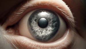
Iris – Colored pigmented surface which excludes the passage of light except through the pupil
Cornea – Transparent dome shaped portion of the eye, that covers the iris acting like a window which emits light into the eyes
Aqueous humor – Transparent fluid that nourishes the lens and the epithelial cells
Lens – Transparent structure of the eye that changes shape to focus incoming light from near and far
Ciliary muscle – Smooth muscle to permit the lens to become more convex
Choroid – Vascular membrane containing pigmented cells
Optic Nerve – Cranial nerve which carries electrical impulses to the brain for interpretation
Fovea – Contains cones and affords the most acute vision
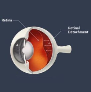
Retina – Stimulation by light which initiates reaction, where the impulses are transmitted to the brain, producing the sensation of vision
Macula – Located in the central retina. Color sensitive rods and central point of the sharpest vision
Vitreous humor – Transparent gelatinous substance filling the eyeball behind the lens
Sclera – Dense white fibrous membrane, forms external covering of eyeball
Ciliary Body / Ciliary Zonules – Holds the lens in place; stretches and loosens
