Although glaucoma cannot be cured, there are treatments available that can effectively manage the condition. Glaucoma treatments are focused on reducing the pressure within the eye (intraocular pressure – IOP) and maintaining a pressure that will reduce the likelihood of further damage to the optic nerve and loss of vision. Often, the vision loss from glaucoma is irreversible.
Glaucoma is often the result of increased pressure inside a person’s eye (intraocular pressure – IOP) caused by a buildup of fluid that leads to vision problems and damage to important structures within the eye, primarily the optic nerve. Glaucoma can develop in one or both eyes. In rare cases, some people that have pressure within a normal range can also develop glaucoma (normal tension glaucoma). Increased eye pressure does not necessarily mean that you have glaucoma.
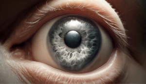
Glaucoma is characterized by the build up of pressure within the eye causing damage to the optic nerve.
Although glaucoma cannot be cured, there are glaucoma treatments available that can effectively manage the condition. In most cases, glaucoma can be successfully treated and managed and if treatment is started early, the majority of people will not experience significant vision loss.
Glaucoma treatment options may include eye drops (topical), oral medications, surgery involving lasers, drug implants, traditional surgical options or a combination approach.[1, 2] The type of treatment will be determined by a number of factors, including the type of glaucoma, cause of glaucoma, response to previous treatment and an individual’s ability to tolerate treatment side-effects.
Glaucoma Treatments and Medications
Glaucoma can be treated with drugs of various classes (types) or a combination of more than one of the following, including prostaglandin analogues, beta blockers, carbonic anhydrase inhibitors, adrenergic agonist, miotics, and hyperosmotic agents. [1, 3] The name of each class of drug describes the action it has on different components of the body (proteins, hormones, etc.). These classes of drugs either increase the fluid flowing out of the eye or reduce the amount of fluid produced in the eye, both providing options to reduce pressure build-up in the eye.
PROSTAGLANDIN ANALOGS
Prostaglandin analogs (PGAs) are the most common class of prescribed eye drops for glaucoma. A prostaglandin is a type of blood protein that is naturally produced by the body and it has the ability to lower intraocular pressure, among other things. The word ‘analog’ indicates that the drug is similar or comparable to the naturally-occurring substance but has a slightly different chemical composition.
PGAs are used to treat open-angle glaucoma because at certain doses they increase the flow of aqueous humor from the eye, reducing pressure inside the eye. Aqueous humor is the fluid made continuously in the eye. PGAs work by relaxing the muscles of the interior of the eye to increase outflow. These eye drops usually cause minimal side effects because they are prescribed at very low concentrations. Some people do experience some eye irritation or redness and darkening of the iris, darkening of eyelashes or eyelid skin, and blurred vision. PGAs are normally prescribed to be used once-a-day.
Some common PGAs include:
- Travatan® (generic name: travoprost)
- Xalatan® (generic name: latanoprost)
- Lumigan® (generic name: bimatoprost)
- Zioptan® (generic name: tafluprost)
CARBONIC ANHYDRASE INHIBITORS
Carbonic anhydrase inhibitors (CAIs) are substances that suppress the action of the enzyme carbonic anhydrase. They are available in eye drops or in pill form. Carbonic anhydrase normally helps to regulate the body’s pH and fluid levels. By suppressing the enzyme, fluid levels in the body are decreased, including the amount of fluid produced by the eye, reducing pressure.
It is important to note that CAIs are sulfonamides so if you have an allergy to sulfa antibiotics, the same types of adverse reactions can occur with CAIs. Also, people taking aspirin in high doses have experienced adverse drug interactions with CAIs.
Some common CAIs include:
- Trusopt® (generic name: dorzolamide hydrochloride) – eye drops
- Azopt® (generic name: brinzolamide) – eye drops
- Diamox®, AK-Zol (generic name: acetazolamide) – oral, pill form
- Diamox® Sequels (generic name: acetazolamide sustained-release) – oral, pill form
- Neptazane®, GlaucTabs (generic name: methazolamide) – oral, pill form
BETA BLOCKERS
Beta blockers are a beta adrenergic receptor agonist, which means that it reduces the pressure in the eye by decreasing the amount of aqueous humor the eye produces. They work by blocking a specific type of nerve endings within the part of the eye that produces fluid (ciliary epithelium), causing a reduction in fluid produced. Beta blockers are classified into two general classes, nonselective and selective. Nonselective beta blockers tend to have more side effects because they act on receptors throughout the body, however the nonselective type has been shown to be more effective at lowering eye pressure.
The eye pressure-lowering effect is slightly less with selective beta blockers. They often require dosing at least twice daily to maintain the pressure lowering effects.
Beta blockers include:
- levobunolol (Betagan®, AKBeta®) – Nonselective beta blocker
- timolol (Timoptic®) – Nonselective beta blocker
- timolol gel (Timoptic® XE) – Nonselective beta blocker
- betaxolol (Betoptic®, Betoptic S®) – Selective beta blocker
Beta blockers also have the ability to decrease someone’s heart rate, so it is important to know that these drugs may cause adverse side effects for those with a medical history of cardiac disease, asthma or slow heart rate (bradycardia).
ALPHA ADRENERGIC AGONISTS
Alpha adrenergic drugs lower eye pressure through alpha adrenergic stimulation, meaning it decreases aqueous humor production by causing constriction in the blood vessels. Possible side effects include an irregular heart rate, high blood pressure, fatigue, red, itchy or swollen eyes, and dry mouth.
Alpha adrenergic agonists include:
- apraclonidine (Iopidine®);
- brimonidine (Alphagan®, Alphagan® P); and
- dipivefrin (Propine®)
MIOTICS
Miotics, also referred to as cholinergic agonists, are eye drops that increase the amount of fluid that flows out of the eye in order to reduce pressure. These drops need to be taken several times per day and some people experience headache, eye ache, smaller pupils, possible blurred or dim vision, and nearsightedness.
COMBINATION GLAUCOMA TREATMENTS
There are several eye drops available that combine the actions of two drugs from different categories. Many people prefer this because they are able to only put one drop in their eye, rather than two or more. Also, it is thought that fewer drops may be healthier for the eyes because it limits the amount of preservative coming into contact with the eye.
Combination medications include:
- Combigan® (Combines a beta-blocker and an alpha-agonist)
- Cosopt® (Combines a beta-blocker and a CAI)
- DuoTrav® (Combines a prostaglandin analog and a beta-blocker)
- Xalacom® (Combines a prostaglandin analog and a beta-blocker)
What you should know about your glaucoma treatment medication?
Adhering to the treatment plan prescribed by your ophthalmologist, including taking medications exactly as directed and attending regular follow-up, is key to preventing optic nerve damage and vision loss. To ensure that you allow the glaucoma medication the best chance to work as intended, it is important to understand a few things about the medication. You can discuss these with your ophthalmologist or pharmacist.
- Know the name of your medication
- Know the dose (how much, how often, when?)
- Know the route of administration (how to take it; as a drop in the eye?, as a pill to swallow?, with meals?)
- Know what you should do if you accidentally miss taking a dose as prescribed (take the next dose as scheduled? Take an extra dose?)
- Know how your medication should be stored (does temperature or exposure to light matter?)
- Know if you can take it with other medications, including non-prescription or natural products. (Can I use my glaucoma eye drops with natural tears? Can I still take ginkgo biloba while taking glaucoma medication?)
- What side-effects can you expect, and when should you seek medical attention for particular side-effects?
WHY DO GLAUCOMA MEDICATIONS HAVE DIFFERENT COLORED BOTTLE TOPS?
Some people take more than one glaucoma medication and it is important to take the correct medication in a specific order. Also, some people receiving glaucoma treatment have already experienced some vision loss. For these reasons, physicians, regulatory agencies and drug manufacturers have agreed that making the bottle top of each type of medication a specific color would be helpful and reduce medication errors.
- Prostaglandin analogues have turquoise bottle caps.
- Carbonic anhydrase inhibitors have orange bottle caps.
- Alpha-adrenergic agonists have purple bottle caps.
- Beta-adrenergic blockers have yellow or blue bottle caps.
Laser and surgical treatments for glaucoma
Selective laser trabeculoplasty (SLT) is a newer and commonly used procedure that can be performed in the ophthalmologist’s office. A laser is used to create new drainage pathways in the eye, allowing fluid to drain more effectively, reducing pressure. Lasers do not burn holes through the eye, they make precise and subtle changes to the drainage system.
Selective laser trabeculoplasty reduces pressure by creating pathways for fluid to drain from the eye.
Before the procedure the ophthalmologist will put numbing drops on the eye, so the patient will not feel any discomfort. They may also put additional drops in the eye to reduce the size of the pupil and put a special contact lens on the eye to direct the laser to very specific areas. During the procedure people may see some short flashes of light, but do not feel anything.
Often the ophthalmologist will create approximately 50 new drainage pathways in an area that includes only half the eye. People may have to return for another visit to have the procedure completed on the other half of their eye.
Although the procedure only takes less than 15 minutes, ophthalmologists usually require patients to stay at the office for a couple of hours to monitor pressure changes in the eye.
Many people that have SLT will still be required to use prescription eye drops to manage their glaucoma. The term ‘selective’ means that this procedure leaves portions of the drainage system (trabecular meshwork) intact, which is important because unlike types of previous laser surgery, SLT can be safely repeated, if needed.
Other laser treatments are available, including argon laser trabeculoplasty (ALT) and micropulse laser trabeculoplasty (MLT). ALT was the first laser trabeculoplasty procedure performed using a thermal (heat) laser which can cause more scarring to the draining pathways than SLT. Although ALT has been shown to reduce pressure in nearly 75% of people, it can be performed only two to three times in each eye over a lifetime due to scar tissue formation. MLT pulses laser energy to the eye tissues in small increments in an effort to reduce the amount of energy delivered to the eye tissues.
LASER PERIPHERAL IRIDOTOMY
Laser peripheral iridotomy is a procedure that uses a laser to create an opening in the iris (the colored part of the eye) enabling fluid to leave the eye more easily, reducing pressure within the eye.
This procedure is most commonly performed on people that have narrow angles (narrow drainage pathways) or their ophthalmologist believes that their angles are more likely to close.
Similar to SLT, numbing drops and a special contact lens are used. The laser will make one or two holes in the iris and the patient may see short flashes of light but will not feel any discomfort.
FILTERING SURGERY (TRABECULECTOMY)
During a trabeculectomy a surgeon creates an opening in the sclera (white part of the eye) and removes part of the trabecular meshwork, the area responsible for draining the aqueous humor from the eye. During the procedure, a flap is created that allows fluid to escape, while maintaining a level of pressure required to keep the eye inflated. A ‘bleb’ is a small bubble of fluid that forms over the opening to indicate that fluid is draining.
Nearly half of people that have filtering surgery no longer require glaucoma medications, while 35-40% still need medication, but their glaucoma is easier to manage.
DRAINAGE TUBES
There are different devices that have been developed to help improve drainage of aqueous humor and manage eye pressure. These drainage devices are made of a small silicone tube connected to one or more plates that are sutured to the surface of the eye. These plates collect fluid and then allow it to be absorbed by other eye tissues.
TUBE-SHUNT SURGERY
Tube-shunt surgery involves placing a flexible plastic tube with an attached silicone drainage pouch in the eye to help drain fluid from the eye. This type of surgery is usually performed after a trabeculectomy has failed. This type of surgery may also be performed initially in people that already have, or are more likely to form, scar tissue in the eye.
MINIMALLY INVASIVE GLAUCOMA SURGERY (MIGS)
MIGS procedures include many of the traditional procedures but use microscopic-sized equipment and tiny incisions. MIGS procedures reduce the incidence of complications but may be less effective for some people. Cataract surgery may also be combined with a MIGS procedure.
MIGS categories include:
- Miniaturized versions of trabeculectomy
- Trabecular bypass operations
- Totally internal or suprachoroidal shunts
- Milder, gentler versions of laser photocoagulation
CATARACT SURGERY
In some cases, cataract surgery alone, replacing the eye’s natural lens with an artificial one (intraocular lens – IOL) have been shown to effectively lower eye pressure in people with open-angle glaucoma patients and narrow-angle glaucoma.
References:
[1] Yadav KS, Rajpurohit R, Sharma S. Glaucoma: Current treatment and impact of advanced drug delivery systems. Life Sci. 2019 Feb 21. pii: S0024-3205(19)30119-5. https://www.ncbi.nlm.nih.gov/pubmed/30797820
[2] McAlinden C. Selective laser trabeculoplasty (SLT) vs other treatment modalities for glaucoma: systematic review, Eye. 28 (2014) 249 https://www.ncbi.nlm.nih.gov/pubmed/24310236
[3] Weinreb RN, Aung T, Medeiros FA. The pathophysiology and treatment of glaucoma: a review. JAMA. 2014 May 14;311(18):1901-11 https://www.ncbi.nlm.nih.gov/pubmed/24825645